About DECI
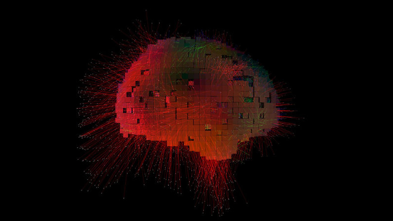
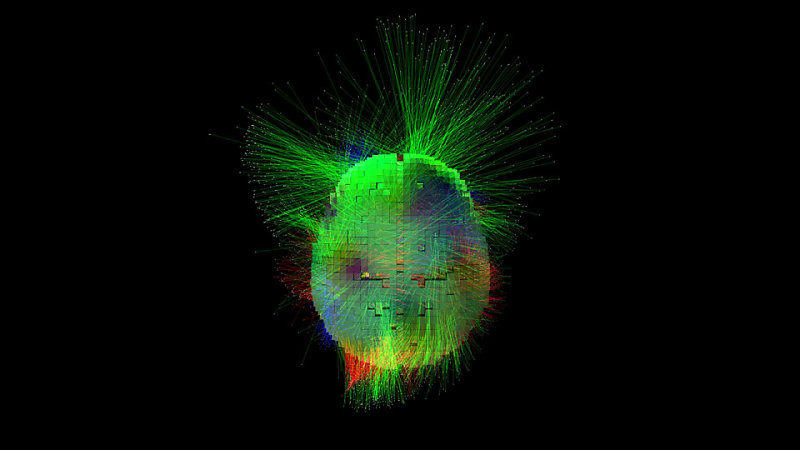
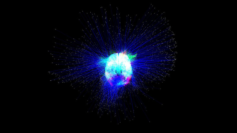
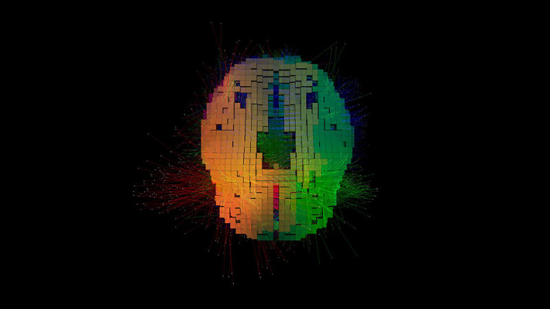
Dynamic Electrical Cortical Imaging (DECI) is a safe, non-invasive, affordable and portable alternative to existing imaging tools such as fMRI, MRI and PET. Unlike, the existing technologies, DECI captures and localizes the electrical activity of the cerebral cortex as it is occurring in real-time and displays it in the form of 3D motion pictures.
HOW DECI WORKS: DECI captures and displays the three-dimensional electrical activity of the brain in real-time using conventional off-the-shelf electroencephalographic (EEG) equipment. Like an EEG, this technology is inherently safe and non-invasive, merely requiring the contact of electrodes to a person’s scalp. However, unlike conventional EEG, whose waves are pale reflections of the underlying complexities within the brain, DECI, through the use of a high-level mathematical transformation known as an inverse solution, produces vivid representations of moving electrical fields. The output of the software includes a stream of images on a screen. Using colour and lines DECI depicts the locations of the generators of the wave-forms seen on an EEG.
BACKGROUND: The human cerebral cortex is the convoluted outer surface of the brain, commonly known as ‘the gray matter’. It is responsible for many of the higher functions of the mind including those associated with thought, action, emotion and sensation. The cerebral cortex, largely due to its complexity, is also susceptible to a large number of disorders and diseases. Many of these diseases lack proper objective diagnostic tests or currently require expensive medical equipment to diagnose them.
Often, in order to diagnose diseases of the brain 3D localization and imaging is required. There are a number of existing technologies that have been utilized that accomplish this including Functional Magnetic Resonance Imaging (fMRI). Each of the existing technologies has its strengths and weaknesses.
5 Simple Advantages of DECI over existing technologies:
- DECI can localize the rapid changes in electrical activity in the human cerebral cortex in three-dimensions in real-time. Thus, with further development and real-time analysis the invention could be used to make an instant diagnosis.
- DECI is very inexpensive when compared to other imaging technologies such as fMRI, CT, MEG and PET.
- DECI is safe and non-invasive.
- DECI is portable. With additional funding, this portability can be further improved, and the entire system can be wireless. This will create a new frontier for psychology and medicine because it will allow for the use of brain imaging to study psychological functions in real life. Presently, brain imaging is only conducted in the stilted environment of laboratories where many important spontaneous emotions and mental processes do not occur.
- DECI can acquire over 2000 images per second. This enables it to capture numerous fine events that are blurred together as one with most competing technologies.
The accuracy of source localization in localizing brain signals
Cerebral Diagnostics is part of an emerging field called source localization. Most new technologies come under fire and over time the best ones stand up to scrutiny. Source localization is gradually gaining respectability. Since its inception 20 years ago there have been many naysayers that have attacked it; but there are also many supporters. If you check PubMed (the massive online data base of medical journal articles), you will find that there are well over 500 hundred articles many of which have appeared in distinguished peer reviewed journals about source localization. Each one had to stand up to scientific scrutiny. The algorithms that find the brain signals in our images were created by mathematicians.
The early algorithms weren’t very accurate in localizing brain signals, especially those deep in the cortex. The new algorithms are getting better and likely to continue to improve. There are multiple scholarly sources of evidence that show that the latest algorithms are at least moderately accurate. One is called evoked potential studies. A second are comparative studies such as the work by Drs. Wennberg and Zumsteg done at the Toronto Western Hospital which found a striking overlap between where PET localized the generator of epileptic activity in the cortex and where source localization placed it.
The third line of evidence in favour of the newer algorithms comes from specialized advanced tests called simulation studies.


